Home
Uncategories
Back Of Neck Anatomy / Neck Sprain Orthoinfo Aaos / The neck muscles, including the sternocleidomastoid and the trapezius, are responsible for the gross motor movement in the muscular system of the head and neck.
Back Of Neck Anatomy / Neck Sprain Orthoinfo Aaos / The neck muscles, including the sternocleidomastoid and the trapezius, are responsible for the gross motor movement in the muscular system of the head and neck.
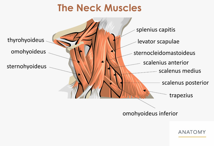
Back Of Neck Anatomy / Neck Sprain Orthoinfo Aaos / The neck muscles, including the sternocleidomastoid and the trapezius, are responsible for the gross motor movement in the muscular system of the head and neck.. The cervical spine, your neck, is a complex structure making up the first region of the spinal column starting immediately below the skull and ending at the first thoracic vertebra. There are two main triangles; The nerves of the head and neck include the most vital and important organs of the nervous system — the brain and spinal cord — as well as the organs of the special senses. It consists of seven vertebrae. The muscles of the neck run from the base of the skull to the upper back and work together to bend the head and assist in breathing.
See more ideas about back pain, spine health, spine problems. The internal jugular veins form the major venous drainage of the head and neck and are deep veins that parallel the common carotid artery. The nose is flanked by two anatomical. These muscles give the sides of the neck their. Occipital neuralgia is caused due to irritation or injury to the occipital nerve.
Neck Back Orthopedic Associates Of Northern California Orthopedic Associates Of Northern California from www.oanc.org The top of the cervical spine connects to the skull, and the bottom connects to the upper back at about shoulder level. The back of the neck is mostly comprised of muscles, as well as the spine. The largest vein in the neck is usually the internal jugular vein, which drains blood from the brain, neck muscles, face and organs of the neck. See more ideas about back pain, spine health, spine problems. Causes of neck pain and how to manage the pain in basic terms, the neck (cervical spine) joins the shoulders and chest to the head. The occipital bone is the only bone in your head that connects with your cervical spine (neck). The occipital bone surrounds a large opening known as the foramen magnum. The internal jugular veins form the major venous drainage of the head and neck and are deep veins that parallel the common carotid artery.
The neck is unique in that it supports the weight of your head (10 to 11 pounds) and allows a variety of head/neck movement, such as turning your head from side to.
Part 7 in an 8 part lecture on skeletal muscle in a flipped human anatomy course taught by wendy riggs. Jugularis posterior) begins in the occipital region and returns the blood from the skin and superficial muscles in the upper and back part of the neck, lying between the splenius and trapezius. Back pain is common and might be caused by a problem with a muscle. The anterior triangle of the neck is made by the anterior border of the sternocleidomastoid muscle, the inferior border of the mandible and the midline of the neck. The neck is essentially a passageway for air, food, liquids, blood, and more to travel between the head and the rest of the body, through structures such as blood vessels, nerves, and lymph nodes, as well as the larynx, trachea, and esophagus. The occipital bone surrounds a large opening known as the foramen magnum. They ascend the posterior aspect of the neck, passing through holes in the transverse processes of the cervical vertebrae (known as foramen transversarium). The external jugular veins descend on either side of the neck, passing over the sternomastoid muscles and beneath the platysma. It consists of seven vertebrae. The majority of these nerves control the functions of the upper extremities and allow you to feel your arms, shoulder, and back of your head. There are two main triangles; Each nerve provides sensation to a specific area of the body called a dermatome. The cervical spine supports the weight and movement of your head and protects the nerves exiting your brain.
Neck anatomy nerves picture there are 8 spinal nerves that originate from the cervical spine. The cervical spine supports the weight and movement of your head and protects the nerves exiting your brain. The internal jugular veins form the major venous drainage of the head and neck and are deep veins that parallel the common carotid artery. It consists of two major. Muscle head anatomy vocal organ diagram female neck anatomy neck wireframe head neck human anatomy head artery anatomy face pharynx vector neck degree head anatomy 3d.
Muscles Of The Neck Teachmeanatomy from teachmeanatomy.info The occipital bone surrounds a large opening known as the foramen magnum. See more ideas about back pain, spine health, spine problems. The neck is connected to the upper back through a series of seven vertebral segments. The majority of these nerves control the functions of the upper extremities and allow you to feel your arms, shoulder, and back of your head. The neck is essentially a passageway for air, food, liquids, blood, and more to travel between the head and the rest of the body, through structures such as blood vessels, nerves, and lymph nodes, as well as the larynx, trachea, and esophagus. It runs from the neck to the upper back. Rarely, neck pain can be a symptom of a more serious problem. Part 7 in an 8 part lecture on skeletal muscle in a flipped human anatomy course taught by wendy riggs.
The majority of these nerves control the functions of the upper extremities and allow you to feel your arms, shoulder, and back of your head.
It runs down the back part of the neck, and opens into the external jugular vein just below the middle of its. In this video, i walk you through a basic approach to drawing the neck and upper back muscles. But don't worry, these triangles are not hard to remember and they are very important for understanding neck anatomy. Rarely, neck pain can be a symptom of a more serious problem. These muscles give the sides of the neck their. The nerves of the head and neck include the most vital and important organs of the nervous system — the brain and spinal cord — as well as the organs of the special senses. Neck anatomy nerves picture there are 8 spinal nerves that originate from the cervical spine. The skull is a strong, bony capsule that rests on the neck and encloses the brain. The neck muscles, including the sternocleidomastoid and the trapezius, are responsible for the gross motor movement in the muscular system of the head and neck. The largest vein in the neck is usually the internal jugular vein, which drains blood from the brain, neck muscles, face and organs of the neck. Occipital neuralgia is caused due to irritation or injury to the occipital nerve. The cervical spine protects the nerves connecting to the brain, allowing the head to move freely while supporting its weight. The internal jugular veins form the major venous drainage of the head and neck and are deep veins that parallel the common carotid artery.
See more ideas about back pain, spine health, spine problems. The skull is a strong, bony capsule that rests on the neck and encloses the brain. The internal jugular vein commences at the jugular foramen, and is the direct continuation of the sigmoid sinus, which is a large vein draining blood from the vein. See anatomy of the head and neck stock video clips. It consists of seven vertebrae.
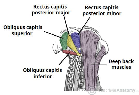
The Back Of Head Neck Muscles Anatomy Page 1 Line 17qq Com from img.17qq.com Anatomy of back of human neck, anatomy of the back and neck, anatomy of the back of the neck, anatomy of the back of the neck muscles, anatomy of the back of your. The majority of these nerves control the functions of the upper extremities and allow you to feel your arms, shoulder, and back of your head. The cervical spine has 7 stacked bones called vertebrae, labeled c1 through c7. The muscles of the neck are present in four main groups. Osteoarthritis also is a common cause of neck pain. The muscles of the neck run from the base of the skull to the upper back and work together to bend the head and assist in breathing. The occipital bone is the only bone in your head that connects with your cervical spine (neck). The suboccipital muscles act to rotate the head and extend the neck.rectus capitis posterior major and rectus capitis posterior minor attach the inferior nuchal line of the occiput to the c2 and c1 vertebrae respectively.obliquus capitis superior also extends from the occiput to c1 while obliquus capitis inferior originates from c2 and.
Neck muscles can be strained from poor posture — whether it's leaning over your computer or hunching over your workbench.
The posterior external jugular vein (v. The anterior, and the posterior, triangles of the neck. It consists of two major. The suboccipital muscles act to rotate the head and extend the neck.rectus capitis posterior major and rectus capitis posterior minor attach the inferior nuchal line of the occiput to the c2 and c1 vertebrae respectively.obliquus capitis superior also extends from the occiput to c1 while obliquus capitis inferior originates from c2 and. Each nerve provides sensation to a specific area of the body called a dermatome. Neck pain is a common complaint. Back pain is common and might be caused by a problem with a muscle. This is a more stylized study and not meant to be entirely cor. An area called the occiput. Sticking out from the middle of your face is your nose, a structure that allows you to smell and breathe. The occipital bone is a bone that covers the back of your head; The muscles of the neck run from the base of the skull to the upper back and work together to bend the head and assist in breathing. The majority of these nerves control the functions of the upper extremities and allow you to feel your arms, shoulder, and back of your head.

0 Comments:
Posting Komentar